Differentiation of confluent hepatic fibrosis and infiltrative hepatocellular carcinoma on MR imaging Jing‑Jing Liu· Meng‑jiao Duan· Meng‑yue Huang · Meng‑na Huang · Meng‑zhu Wang · Yong Zhang· Jing‑liang Cheng Confluent fibrosis (CF) is a benign condition characterized by the development of extensive fibrotic scars in the liver, commonly observed in patients with advanced cirrhosis. Despite its prevalence, the exact cause of CF remains unknown, and its differentiation from malignant tumors, such as hepatocellular carcinoma (HCC), it’s particularly challenging. This study aims to identify key MRI features, considered alone, and combined, that can effectively distinguish CF from infiltrative HCC, thereby facilitating accurate diagnosis and optimal patient management. A retrospective analysis was conducted on patients with chronic liver disease who presented with either focal confluent fibrosis (FCF), confluent fibrosis (CF), or infiltrative hepatocellular carcinoma (HCC). All patients underwent liver MRI using 3 Tesla MRI scanner. Inclusion criteria required patients to have undergone conventional MRI, dynamic contrast-enhanced MRI (DCE-MRI), and diffusion-weighted imaging (DWI) exams within two weeks prior to surgery or pathological biopsy, with pathology confirmation through biopsy or surgery, or observation of stable lesion size for at least one year during follow-up. Forty-eight patients were included for the final analysis. The study cohort comprised 20 CF patients, including 4 (20.0%) males and 16 (80.0%) females, with a mean age of 55 ± 14 years (range: 17 to 80 years). The infiltrative HCC group consisted of 28 patients, with 23 (82.1%) males and 5 (17.9%) females, and a mean age of 54 ± 12 years (range: 34 to 76 years). CF was significantly more prevalent in females (P < 0.001). The distinguishing MRI features between CF and infiltrative HCC were identified. CF patients exhibited more hepatic capsular retraction, hepatic volume loss, and a "nodular surround sign" compared to infiltrative HCC patients (all P < 0.001). Conversely, infiltrative HCC patients demonstrated more hemorrhage, cystic changes, and macrovascular invasion (all P < 0.05). Additionally, CF lesions often displayed no or mild enhancement in the arterial phase of DCE-MRI but showed hyperintensity in the delayed phase, whereas infiltrative HCC lesions typically exhibited marked enhancement in the arterial phase followed by washout in the delayed phase. Furthermore, all cases showed high signal intensity on DWI, with CF lesions demonstrating significantly higher apparent diffusion coefficient (ADC) values compared to infiltrative HCC lesions (P < 0.001). Three MRI categories showed notably diagnostic yield for CF: hepatic volume loss at the lesion site (83.3% of cases), no or mild arterial phase enhancement (85.4%), and hyper-enhancement in the delayed phase (97.9%). Combining these individual MRI categories resulted in even higher diagnostic accuracy. Combinations involving hepatic volume loss, no or mild enhancement in the arterial phase, and hyper-enhancement in the delayed phase achieved 100.0% sensitivity for CF diagnosis, with specificity reaching 100.0% when any two or all three categories were positive. The combination yielding the highest diagnostic accuracy (95.8%) included lesions showing no or mild enhancement in the arterial phase or hyper-enhancement in the delayed phase, along with hepatic volume loss. Among the limitations of the study, the use of vasculo-interstitial CM was not reported. Hepatobiliary contrast medium has a significative role for the differential between CF and HCC. Notably, Park et al. emphasized the effectiveness of Gd-EOB-DTPA , reporting an incremental value compared to T2-weighted imaging for the determination of outer margin, shape and extent of infiltrative HCC and distribution and extent of satellite nodules. They also found that CF tended to have a higher lesion-to-liver signal intensity ratio than that of infiltrative HCC on hepatobiliary phase imaging. In conclusion, CF patients often exhibit hepatic volume loss, hepatic capsular retraction, no or mild enhancement in the arterial phase, hyper-enhancement in the delayed phase, higher ADC values and higher lesion to liver signal intensity ratio on hepatobiliary phase. If these three features are present (e.g. hepatic volume loss, arterial phase enhancement, and delayed phase hyper-enhancement) diagnostic yielded rise to 96% for CF diagnosis. |
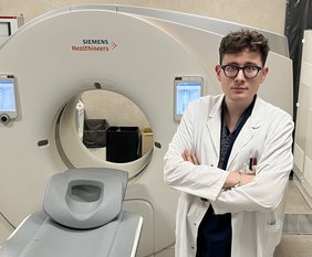
Dr. Saletti Carlos Leon is a first-year radiology resident on the “Sapienza, University of Rome” training scheme in Italy. He completed his undergraduate medical degree at “Sapienza, University of Rome” in 2023. He joined the Medical Imaging Department in the same year where he is undertaking training in diagnostic and interventional radiology, with particular interest towards abdominal imaging. Comments may be sent to: saletti.leon@gmail.com |

