Contrast-enhanced US of High‑Risk Indeterminate Focal Liver Observations Categorized as LR‑4 or LR‑M at CT/MRI CEUSLI‑RADS Trial Group; Lyshchik A, Escalante CKY, et al. Radiology. 2025;314(1):e240916. DOI:10.1148/radiol.240916 Introduction Differentiating focal liver lesions in at-risk patients remains a cornerstone of hepatobiliary imaging, particularly in the setting of cirrhosis and chronic hepatitis B. Although multiphasic CT and MRI, along with LI-RADS classification, provide structured pathways toward diagnosing hepatocellular carcinoma (HCC), indeterminate categories such as LR-4 ("probably HCC") and LR-M ("probably malignant, not specific for HCC") continue to present clinical uncertainty [1,2]. These ambiguous findings often lead to delayed diagnoses, biopsies, or prolonged follow-up. In recent years, contrast-enhanced ultrasound (CEUS) has gained recognition as a valuable adjunct for liver lesion characterization. Its real-time imaging capability, lack of ionizing radiation, and safety in renal impairment make it an appealing modality, especially in complex or equivocal cases [3]. The CEUSLI-RADS study offers timely insight into how CEUS can enhance decision-making in cases where cross-sectional imaging falls short. Study Methods This international, multicenter, prospective trial enrolled 319 patients with chronic liver disease who had indeterminate focal liver lesions categorized as LR‑4 or LR‑M on CT or MRI. All underwent CEUS by experienced operators, blinded to prior imaging and clinical data. The lesions were reclassified according to CEUS LI-RADS (version 2017), and final diagnoses were confirmed via histopathology or composite clinical and imaging follow-up over 24 months [1]. Discussion of Results The study found that CEUS led to reclassification in 33% of cases: 30% were upgraded to LR‑5 (definite HCC), with 94% of these confirmed as HCC, while 10% were downgraded to benign categories (LR-1 or LR-2), demonstrating a 96% concordance rate [1]. These findings align with earlier data from Schellhaas et al., who reported an 84% diagnostic accuracy for CEUS LI-RADS in a similar setting [4]. A meta-analysis by Wu et al. further supports CEUS's comparable performance to CT/MRI in terms of sensitivity and specificity for HCC [5]. Notably, CEUS proved especially helpful in clarifying vascular profiles. In many cases where CT/MRI showed atypical arterial enhancement or delayed washout, CEUS provided classical vascular behavior—prompt wash-in and late mild washout—facilitating confident reclassification. Previous research has similarly highlighted CEUS’s capacity to detect hypervascularity in small or subcapsular lesions not easily visualized on MRI, especially in patients with renal impairment or contrast allergies [6,7]. These findings underscore the complementary nature of CEUS. Rather than replacing CT or MRI, CEUS may serve as a real-time problem-solving tool when standard imaging leaves diagnostic gaps. | Strengths and Limitations The study's strengths include its prospective design, multicenter approach, standardized protocols, and blinded interpretations. Including both LR‑4 and LR‑M categories mirrors real-world diagnostic uncertainty and enhances clinical relevance. However, several limitations are acknowledged. Not all lesions were confirmed histologically, and some relied on follow-up rather than biopsy, introducing potential verification bias. Additionally, CEUS remains operator-dependent and may be limited in obese patients or those with poor acoustic windows [8]. The washout criteria used for CEUS LI-RADS also remain a subject of ongoing debate, particularly in differentiating LR-M lesions [9]. Future Perspectives and ESGAR Relevance The findings strongly support the integration of CEUS into diagnostic algorithms for indeterminate liver lesions, particularly in cases where CT or MRI results are equivocal. As artificial intelligence, fusion imaging, and contrast technologies evolve, CEUS may become even more precise and standardized [10]. For ESGAR members, especially those practicing in high-volume or resource-constrained settings, this study emphasizes the value of CEUS in streamlining care and improving diagnostic confidence. It reinforces the shift toward multimodality imaging and tailored, patient-centered diagnostic strategies in hepatobiliary radiology. References
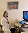
Junior reviewer Dovile Volodkoviciene is currently in her final year of radiology residency at Vilnius University Hospital Santaros Clinics. Having previously completed a residency in internal medicine, radiology represents her second specialization. Dovile is deeply engaged in gastrointestinal imaging, with a particular focus on hepatobiliary radiology. Comments may be sent to: dovile.janulynaite(at)santa.lt |

